In a new study, UK DRI Emerging Leader Dr Maura Malpetti and Harry Crook (UK DRI at Cambridge) developed a method of standardising brain imaging results for PSP and Alzheimer’s. The research, published in the European Journal of Nuclear Medicine and Molecular Imaging, paves the way for larger studies and more accurate tracking of brain inflammation in neurodegenerative diseases, which could lead to new therapies.
What was the challenge?
Microglia are the brain’s resident immune cells, acting as the primary defence to protect the brain from damage. But when these cells stay active for too long, they can cause inflammation in the brain – known as neuroinflammation. Research has shown that neuroinflammation plays a key role in diseases like Alzheimer’s and progressive supranuclear palsy (PSP).
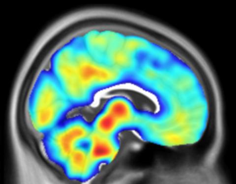
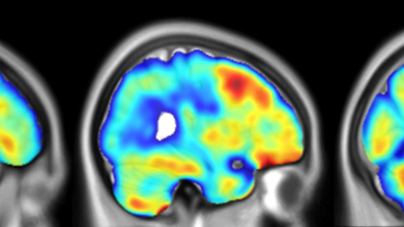
To study inflammation in the living brain, researchers use a type of brain scan called PET imaging. This scan uses special dyes, called radiotracers, that are injected in the blood of participants and attach to a protein called TSPO, which becomes more active when there is inflammation in the brain. Different research centres have developed various TSPO radiotracers, but their results are not always easily comparable. In this study, the team looked at how to make results from different TSPO tracers more consistent and comparable across research centres.
What did the team do and what did they find?
The researchers brought together data from people with PSP who were scanned with two types of radiotracers, and people with Alzheimer’s disease who were scanned with three types, across Cambridge, Munich and Montreal. After applying a method to standardise the imaging data, they found that results from the PSP groups matched up well, but there were more differences in the Alzheimer’s groups depending on the tracer used.
What is the impact?
This research is an important step toward making brain inflammation scans more reliable and standardised, no matter where or how they are done. By unifying the data, scientists will be able to combine results from multiple centres around the world. This will allow for larger studies and more accurate tracking of inflammation in brain diseases, which could help test new treatments and measure how well they work.
This work brings us a step closer to speaking the same scientific language when it comes to imaging brain inflammation. By standardising results across centres and tracers, we can better compare data worldwide and build a clearer picture of neuroinflammation in diseases like Alzheimer’s and PSP.
Emerging Leader