New research led by Dr Johanna Jackson and Prof Paul Matthews (UK DRI at Imperial) in collaboration with researchers at McGill University, reveals how scientists could better preserve human brain tissue for laboratory research. The study, published in Nature Communications, could accelerate the development of treatments for neurodegenerative diseases and inform best practices for how brain banks collect and preserve tissue worldwide.
What was the challenge?
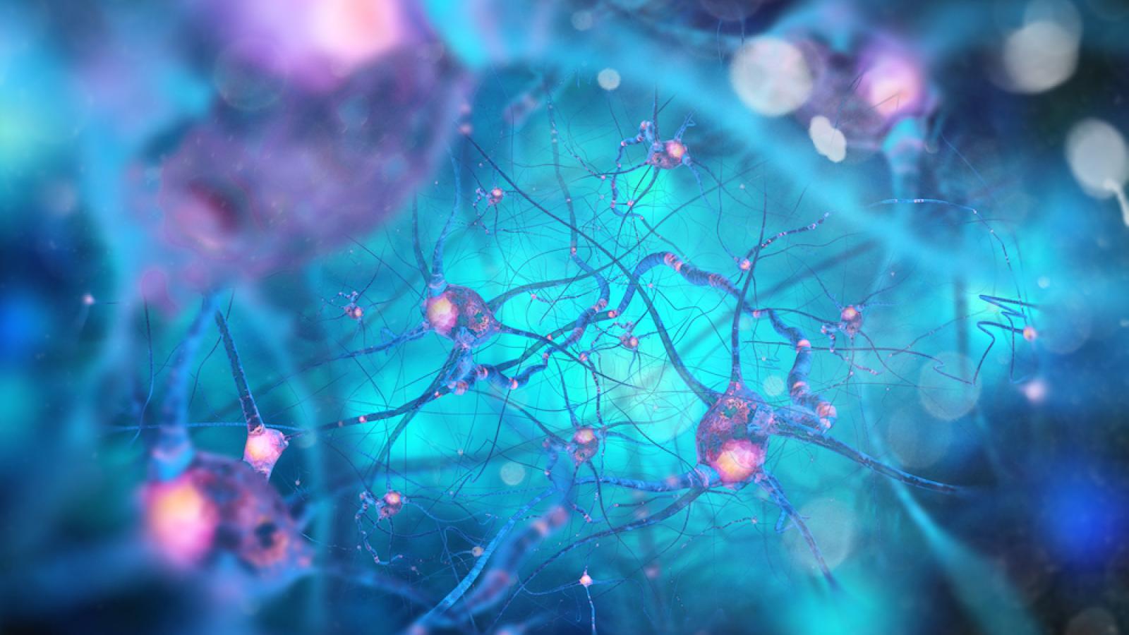
A significant amount of neuroscience research relies on human post-mortem brain tissue, the quality of which deteriorates after death.
The time between death and tissue collection, known as the postmortem interval (PMI), is a key determinant of the quality of brain tissue samples. Previous research confirmed that within just a few hours of death, genes associated with neuronal activity begin to shut down, while immune cells (microglia and astrocytes) respond to the damage and begin clearing dying cells.
The longer the tissue is left before collection, the more its genetic profile diverges from what it looked like in the living brain.
What did the team do and what did they find?
The researchers analysed tissue from McGill in Canada and post-mortem tissue from brain banks in the UK and the Netherlands.
The study compared gene expression in brain tissue extracted immediately from brains during surgery (< 0 hours) and brain tissue with short (~6 hours) and long (~36 hours) post-mortem intervals (PMIs). Researchers confirmed that even a 6-hour postmortem interval caused significant changes in gene activity compared to freshly extracted tissue. The genes driving these deviations were named Brain Artifact Genes (BAGs).
To separate the effects of time and temperature, the team stored fresh tissue at either room temperature (20°C) or in a fridge (4°C) for 6, 24, and 36 hours. Tissue kept at 4°C for 6 hours showed no changes, while just 6 hours at room temperature was enough to trigger them.
Researchers also proved that different brain cell types were affected at different times. After six hours at room temperature, glutamatergic neurons, responsible for memory and learning, were the first to show damage, followed by GABAergic neurons, which regulate mood, sleep and anxiety. At 24 hours, oligodendrocytes, which allow signals to travel efficiently, became the most affected, followed by microglia, the brain’s immune cells.
This work has also shown that the temperature at which the brain is kept may have greater implications than the time before being used in post-mortem transcriptomics studies.
Emerging Leader
What is the impact?
Until now, there was no tool available to systematically measure and account for cellular changes in the brain after death. Using deep learning, the team distilled the broader processing-response program into a compact, predictive signature called Time and Temperature Response genes Underlying Transcriptional Heterogeneity (TTRUTH).
The model assigns a continuous score to individual autopsy samples, estimating the extent to which time and temperature have affected their gene activity data. The tool is freely available to the global research community at brainttruth.openscience.mcgill.ca, allowing brain banks worldwide to better standardise datasets and enhance data interpretation.
Dr Jo Anne Stratton, co-corresponding author, McGill University, said:
"This is an excellent online resource so anyone can upload datasets to this user-friendly open website to identify brain-relevant transcripts susceptible to processing artifacts. Brain autopsies are notoriously time-consuming and unpredictable, and RNA, which degrades rapidly after death, makes interpreting data on gene activity in brain tissue particularly challenging. We hope this resource will help, even just a little, to make better sense of the vast and ever-growing number of brain omics datasets out there.
Prof Paul Matthews, Edmond and Lily Safra Chair at the Department of Brain Sciences at Imperial, and article co-author, said:
“This study addresses an important source of potential bias in the study of post-mortem human tissues with the increasingly widely used single nuclear transcriptomic technologies. In other words, what looks like a biological difference may simply be an artefact of how the tissue was handled after death. What is exciting is that it suggests that the time- and temperature-dependent changes are consistent enough to allow them to be recognised and allowed for in interpretation of the data.”
Dr Jackson concluded:
“Whilst time and temperature are both important, this means that if the brain can be kept cool as soon as possible after death, the quality of the data may be improved. This has implications for post-mortem and brain banking services.”
Reference: Yaqubi, M., Thomas, M., Talbot-Martin, J. et al. Characterising processing conditions that artifactually bias human brain tissue transcriptomes. Nat Commun 17, 2848 (2026). https://doi.org/10.1038/s41467-026-68872-9
Source: Imperial College London