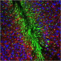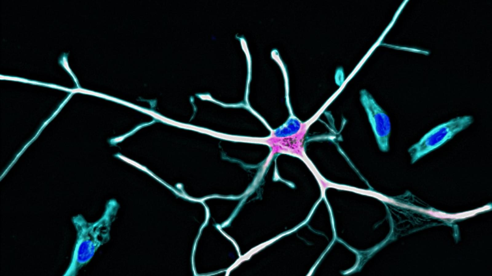
A new study led by Associate Prof Mootaz Salman and Dr Luise Schlotterose, BHF-UK DRI Centre for Vascular Dementia Research (CVDR) at the University of Oxford, has developed a human “TBI-in-a-dish” model that reveals how repeated mild brain injury can leave support cells in the brain persistently altered and trigger damage to neighbouring neurons. Published in Cell Death & Disease, the study recreates repeated mechanical injury in human cells, allowing researchers to follow the cellular consequences of trauma beyond the initial impact.
What was the challenge?
Traumatic brain injury is one of the leading causes of long-term disability, and there's growing evidence that repeated mild head injuries, like concussions, can raise the risk of dementia and other neurodegenerative conditions later in life. There is an urgent need for treatments that can prevent the damage to cells that continues after the initial injury.
What did the team do and what did they find?
The study looked at astrocytes, the star shaped cells that support and protect neurons in the brain. Using human astrocytes grown in the lab, the researchers gave the cells a series of small, carefully controlled injuries to mimic repeated mild head trauma.
The repeatedly injured astrocytes changed in several ways. Their internal structure was reshaped, they built up harmful molecules linked to cell stress, they swelled and shrank, they altered the material around them, and they began storing fat in small droplets. Importantly, these changes suggest that astrocytes retain a form of ‘cellular memory’ of trauma, with the consequences of injury continuing to evolve long after the initial impact.
Crucially, the study also revealed an unexpected mechanism connecting injured astrocytes to subsequent neuronal death. Rather than a classic inflammatory response pathway, the injury altered signalling pathways involving fat molecules. The astrocytes released higher levels of particular fats called diacylglycerols. This in turn switched on a signalling pathway in neurons called PKC-delta. The team found that blocking this pathway partly protected neurons from damage, identifying a potentially druggable mechanism linking traumatic injury to delayed neurodegeneration.
Associate Prof Salman said:
“One of the major challenges in developing treatments for brain injury is translating mechanisms identified experimentally into therapies that are relevant to human disease. Humanised models such as our TBI-in-a-dish platform can help bridge that gap by allowing us to recreate key features of injury in human cells, uncover mechanisms that drive secondary damage and, importantly, test whether these pathways can be therapeutically targeted.
“Our next step is to build increasingly sophisticated human models incorporating different components of the neurovascular unit and use these platforms for higher-throughput drug screening. Ultimately, we want to create a translational pipeline that takes us from understanding mechanism to identifying and testing potential treatments.”
What is the impact?
The “TBI-in-a-dish” platform provides a controllable and scalable human experimental system in which candidate mechanisms can be targeted directly. This opens the door to screening larger libraries of compounds to identify drugs capable of preventing or reversing the secondary damage that follows TBI.
The research was supported by the Medical Research Council, British Heart Foundation, UK Dementia Research Institute and BHF Oxford Centre of Research Excellence, with additional fellowship support for Dr Schlotterose from the Royal Society.
Reference: Schlotterose, L., Struwe, M.A., Scheidig, A.J. et al. From injury to degeneration: delayed lipid-mediated neurotoxicity in a human astrocyte traumatic brain injury in-a-dish model. Cell Death Dis (2026). https://doi.org/10.1038/s41419-026-09217-8